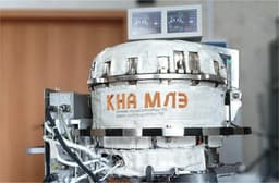
At Peter the Great St. Petersburg Polytechnic University (SPbPU), an innovative approach to three-dimensional internal visualization has been developed. This method detects inhomogeneities in the structure of various objects 3-5 times better than magnetic resonance imaging (MRI) and X-ray. This was reported in the press service of the university.
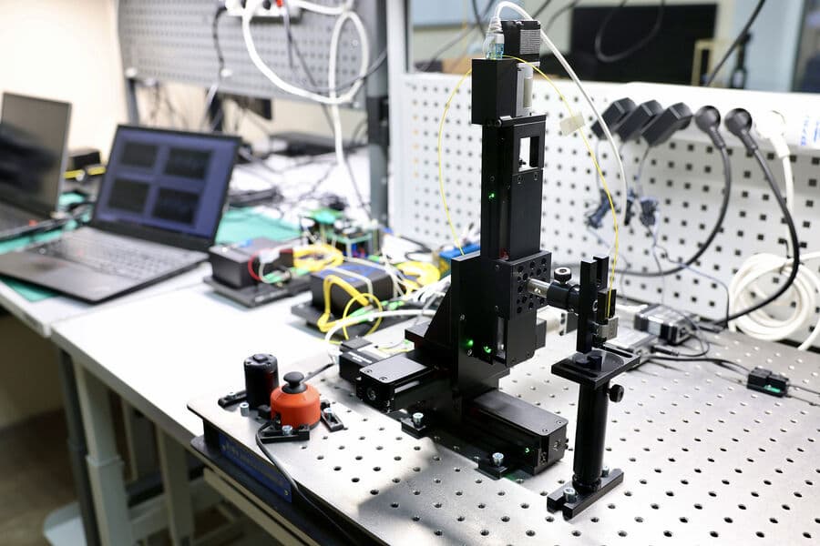
Scientists have proposed a comprehensive improvement of existing optical coherence tomography technologies for use in medicine and industry. They optimized the setup and signal processing methods.
Our method will allow us to check and reject parts of optical and optoelectronic systems at an early stage of production, which will reduce costs. In biology, the new approach will allow monitoring the growth of cells in living organisms in real time or visualizing the delivery and action of drugs inside tissues.
Earlierwww1.ru reported that Moscowis creating a unified network of diagnostic medical devices - from MRI to mammography machines.
Read materials on the topic:
Nanodroplets for accurate diagnostics using MRI and ultrasound developed at Skoltech
Patients with depression can now be identified using MRI
Russian scientists have created a neural network to improve the diagnosis and removal of tumors
Now on home

Start of deliveries scheduled for 2027

Over 51,000 new motorcycles were sold in Russia in 2025

The car will take at least a year to assemble

The application's audience has reached 20 million users

The model will be included in the list of cars for taxis, price - from 2.25 million rubles

All parking lots of the "Administrator of the Moscow Parking Space" are connected to the service

The cars will be supplied to the Moscow Transport Service Directorate
Deliveries to India may begin in 2028
The technology provides automated search for all types of defects in power units
The plane flew 500 km, accelerating to 425 km/h

The plant stated that the information about the termination of purchases for models 6 and 8 is not true
Scientists are using the "Ekran-M" installation