Scientists at Penza State University have developed a web application for diagnosing coronavirus pneumonia using chest X-rays without the need for computed tomography.
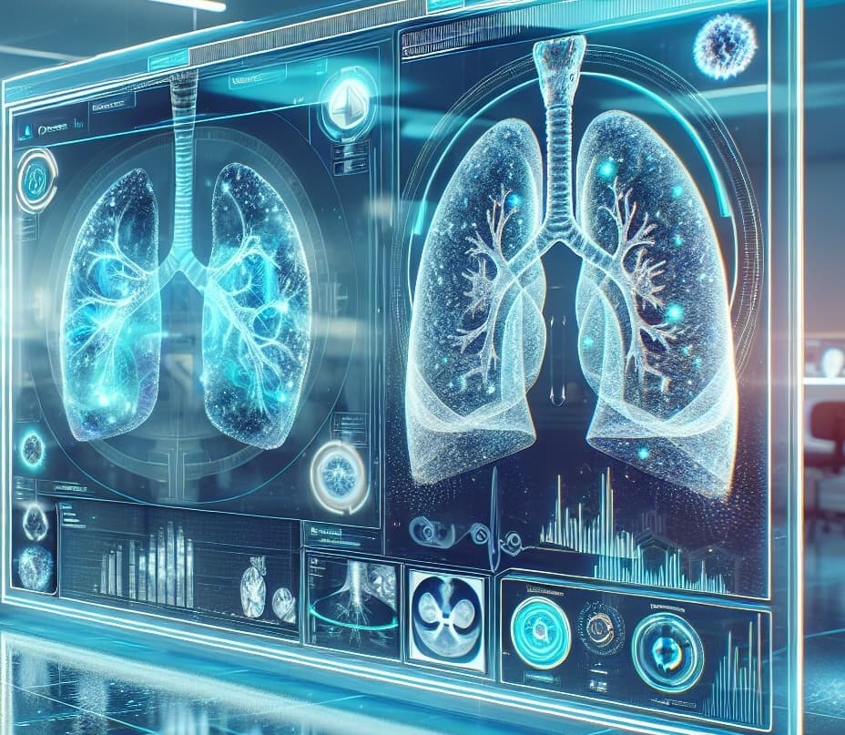
The new system helps radiologists more accurately identify signs of viral pneumonia caused by COVID-19 on X-ray images. The neural network, trained on 1240 X-ray images, can analyze data and determine the degree of lung damage. The application helps reduce the use of expensive and not always available tomography, providing rapid diagnosis with an accuracy of 90-95%.
The developed method allows for the effective detection of the disease at an early stage and is an important tool for screening COVID-19, especially in a pandemic. The program significantly improves the diagnostic process and helps doctors make decisions as quickly as possible.
Read materials on the topic:
AI on guard of health: a project to analyze tomography for cancer diagnosis has started at NSU
Комментарии